Department of Laboratory Medicine and Pathology, University of Minnesota Medical Center, Fairview, Minneapolis, MN
|
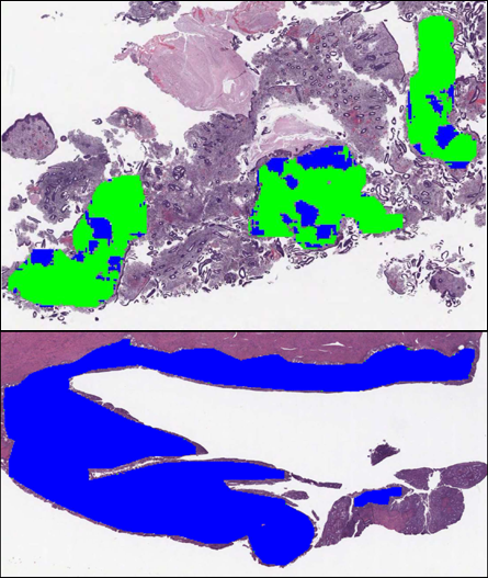
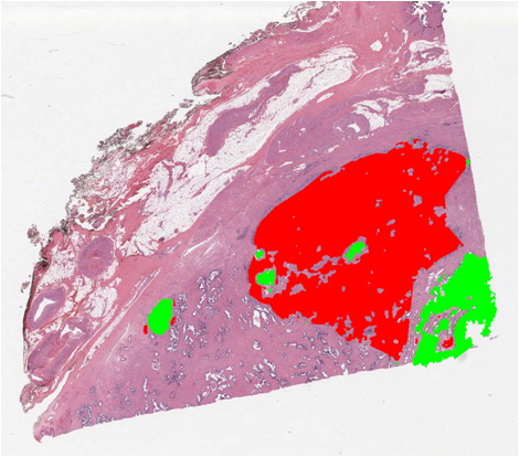
Early diagnosis is quintessential for the effective treatment of disease. For a surgical pathologist, the most time-consuming aspect of the diagnostic process involves arduously scrutinizing tissue slides under a microscope for the evidence of disease. As a result, even a skilled pathologist is able to diagnose only a few patients every day. Our goal is to expedite this process through computer-assisted diagnosis. As a significant step in this direction, our goal is to develop novel image processing and machine learning algorithms tailored toward automated and semi-automated cancer diagnosis. Preliminary results on endometrioid and adenocarcinoma have produced favorable results and overwhelming positive feedback from the community. Specifically the goal of this project is to reduce the time spent by pathologists on each tissue slide. To achieve this we focus on three main sub-tasks. The first step is automatic segmentation in which we aim to direct the attention of the pathologist towards the regions which are likely to be cancerous with minimal false negative (miss) rate and false positive (false alarm) rate. Typical outputs of this step are `high confidence cancer regions', 'low confidence cancer regions' and 'high confidence benign regions' determined automatically by the segmentation step. Based on the segmented regions, our second step is to identify the disease associated with the candidate malign regions. This process entails building appearance based modeling for capturing the unique visual cues that a pathologist uses to identify a disease. There is a significant contribution of low-level image features and high-level computer vision/ machine learning algorithms. As a final step, we plan on using the data-driven computational models of the different pathological conditions to interpret and confirm more complicated medical conditions. Our specific objective is for experts to use this framework as a tool for discovering and developing a deeper insight into these medical conditions. For example, we are interested in determining the "fine-line" or boundaries between the different stages of cancer. Projects
Publications Ravishankar Sivalingam, Guruprasad Somasundaram, Xinyan Li, Alesia Kaplan, Jonathan Henriksen, Arindam Banerjee, Vassilios Morellas, Nikolaos Papanikolopoulos, and Alexander Truskinovsky To appear in the Annual Meeting of the United States & Canadian Academy of Pathology (USCAP), 2012. Ravishankar Sivalingam, Guruprasad Somasundaram, Aravind Ragipindi, Arindam Banerjee, Vassilios Morellas, Nikolaos Papanikolopoulos, and Alexander Truskinovsky Annual Meeting of the United States & Canadian Academy of Pathology (USCAP), 2011. People
Faculty Graduate Students  Aravind Ragipindi Aravind Ragipindi | |||||||||
|
|